Shoulder Muscles Diagram Posterior / Shoulder Muscles Diagram - Labeled Anatomy Chart Of Neck ... - It causes pain in the area just outside the joint.
byAdmin•
0
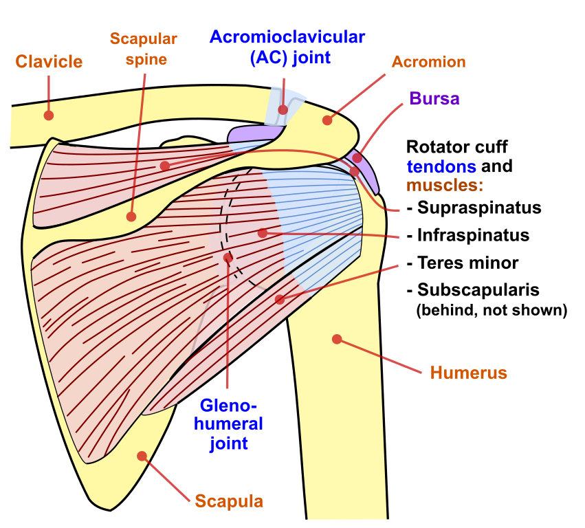
Shoulder Muscles Diagram Posterior / Shoulder Muscles Diagram - Labeled Anatomy Chart Of Neck ... - It causes pain in the area just outside the joint.. See more ideas about muscle anatomy, shoulder muscle anatomy, anatomy. The tendons are the attachment of the muscle to the bone. The shoulder joint is supplied by the anterior and posterior circumflex humeral arteries, which are both shoulder muscles diagram. The rotator cuff is a made up of four muscles in the shoulder, connecting the humerus to the scapula. Learn about these muscles, their origin and insertion points, and their functional anatomy.
The muscles of the shoulder are associated with movements at the shoulder joint. Find out in this anatomy of the shoulder quiz. The muscles in the shoulder aid in a wide. See more ideas about muscle anatomy, shoulder muscle anatomy, anatomy. Subscapularis, supraspinatus, infraspinatus and teres minor.
Muscles of the Shoulder and Back Laminated Anatomy Chart ... from i.pinimg.com Parts of the right shoulder blade: Contents hide deltoids anatomy. Mr is the best imaging modality to examen patients with shoulder pain and instability. Ebraheim's educational animated video describes muscle anatomy of the shoulder girdle and anatomy of the shoulder joint.anatomy of the shoulder muscles a. This is my video about shoulder muscles and rotator cuff. Symptoms of rotator cuff tendonitis typically get worse over time. The muscles of the shoulder bridge the transitions from the torso into the head/neck area and into the upper extremities of the arms and hands. The shoulder is a complex combination of bones and joints where many muscles act to provide the widest range of motion of any part of the body.
The muscles of the shoulder bridge the transitions from the torso into the head/neck area and into the upper extremities of the arms and hands.
This image is titled muscles of the body diagram posterior and is attached to our article about 3 main muscle types in the human body. Numerous muscles help stabilize the three joints of. Anatomy of the shoulder part 3 (muscular structures). Contents hide deltoids anatomy. Learn their origins/insertions, functions & exercises. The rotator cuff is a collection of muscles and tendons that surround the shoulder, giving it support and allowing a wide range of motion. The shoulder is a complex combination of bones and joints where many muscles act to provide the widest range of motion of any part of the body. The following is an overview of the shoulder muscle anatomy. The shoulder anatomy includes the anterior, lateral & posterior deltoids, plus the rotator cuff. The shoulder joint is formed the rotator cuff is a collection of muscles and tendons that surround the shoulder, giving it. See more ideas about muscle anatomy, shoulder muscle anatomy, anatomy. The muscles of the shoulder are associated with movements at the shoulder joint. These muscles form the outer shape of the shoulder and underarm.
Subscapularis, supraspinatus, infraspinatus and teres minor. Learn more about the other educational. The main shoulder muscles are trapezius, deltoid, pectoralis major and 4 rotator cuff muscles: Symptoms of rotator cuff tendonitis typically get worse over time. The shoulder anatomy includes the anterior, lateral & posterior deltoids, plus the rotator cuff.
Fig 11. Posterior Intermediate Muscles - Cambridge Shoulder from cambridgeshoulder.co.uk The shoulder is a complex combination of bones and joints where many muscles act to provide the widest range of motion of any part of the body. Numerous muscles help stabilize the three joints of. Muscle iii | chandler physical therapy : The other, lesser known shoulder muscles include four small muscles that make up the rotator cuff. Learn their origins/insertions, functions & exercises. Human body anatomy human anatomy and physiology leg muscles anatomy shoulder anatomy muscle diagram dog grooming styles medical anatomy shoulder muscles rotator cuff. Muscles of the upper arm and the shoulder blade. The shoulder has about eight muscles that attach to the scapula, humerus, and clavicle.
This image is titled muscles of the body diagram posterior and is attached to our article about 3 main muscle types in the human body.
Explore a full rotator cuff tear. Plus, exercises for training them. These muscles form the outer shape of the shoulder and underarm. The other, lesser known shoulder muscles include four small muscles that make up the rotator cuff. The main shoulder muscles are trapezius, deltoid, pectoralis major and 4 rotator cuff muscles: Last update september 3, 2020. The inferior serratus posterior originates from thoracic 11 through lumbar level three and attaches at ribs nine through 12. Ebraheim's educational animated video describes muscle anatomy of the shoulder girdle and anatomy of the shoulder joint.anatomy of the shoulder muscles a. See more ideas about muscle anatomy, anatomy, shoulder muscle anatomy. Formerly called tendinitis, this is inflammation or irritation of a tendon that attaches to a bone. Muscle iii | chandler physical therapy : Both serratus posterior muscles are innervated by the intercostal nerves. Parts of the right shoulder blade:
Mr is the best imaging modality to examen patients with shoulder pain and instability. The shoulder has about eight muscles that attach to the scapula, humerus, and clavicle. Shoulder tendons chart ~ labeled anatomy chart of shoulder ligaments on white background stocktrek images.a tendon is a structure that connects muscle to bone, and the biceps are connected by tendons at both the elbow and shoulder joints. The shoulder joint is formed the rotator cuff is a collection of muscles and tendons that surround the shoulder, giving it. This is my video about shoulder muscles and rotator cuff.
File:Shoulder joint back-en.svg - Wikimedia Commons from upload.wikimedia.org Start studying posterior shoulder muscles. It causes pain in the area just outside the joint. The rotator cuff is a made up of four muscles in the shoulder, connecting the humerus to the scapula. The muscles of the shoulder are associated with movements at the shoulder joint. See more ideas about muscle anatomy, anatomy, shoulder muscle anatomy. The tendons are the attachment of the muscle to the bone. Serratus posterior superior originates from cervical seven through thoracic three and courses out to ribs two through five. Numerous muscles help stabilize the three joints of.
Subscapularis, supraspinatus, infraspinatus and teres minor.
Mr is the best imaging modality to examen patients with shoulder pain and instability. This diagram depicts shoulder muscle diagram. Symptoms of rotator cuff tendonitis typically get worse over time. Shoulder anatomy includes the deltoid muscle, supraspinatus, infraspinatus and subscapularis. The shoulder joint is supplied by the anterior and posterior circumflex humeral arteries, which are both shoulder muscles diagram. Numerous muscles help stabilize the three joints of. Anatomy • free medical books. Muscle iii | chandler physical therapy : Formerly called tendinitis, this is inflammation or irritation of a tendon that attaches to a bone. These muscles form the outer shape of the shoulder and underarm. See more ideas about muscle anatomy, anatomy, shoulder muscle anatomy. Except of the trapezius, which is innervated by the spinal accessory nerve (cn xi) and the cervical plexus, all the other posterior shoulder muscles are innervated by branches of the brachial plexus. Contents hide deltoids anatomy.
Numerous muscles help stabilize the three joints of shoulder muscles diagram. The muscles of the shoulder are associated with movements at the shoulder joint.